
The Biology of Color
How do cell biologists visualize cells?
Visualization has always been an important component of cell and molecular biology. Antoni van Leeuwenhoek marveled at the diversity of “animalcules”, using a single lens microscope that he designed himself. Since then, microscopy has come a long way, but had still depended on the visible spectrum of light to illuminate cells and structures. That is, until the discovery of Green Fluorescent Protein, or GFP.
GFP was discovered in the Aequorea victoria jellyfish, a small 238 amino acid protein, which glowed green when excited by light in the blue to ultraviolet range. The most exciting discovery that has made GFP the staple of cell biology is its ability to be used as a fluorescent tag for proteins within the cell (1). The GFP protein is so small that its sequence can be glued together with the sequence of another protein, producing a fluorescent version of the protein of interest, without loss of function (as a standard protocol, the functionality of the GFP fusion protein has to be tested for wild type behavior). This enables the protein of interest to be visualized and studied in its natural environment by fluorescence microscopy. The discovery of fluorescent proteins earned Osamu Shimomura, Martin Chalfie and Roger Y. Tsien the Nobel Prize for Chemistry in 2008 (2).
There were issues however with GFP early on – the protein was not stable enough and faded quickly, and it had to be successfully produced at various temperatures that were considerably different from the environment in which the jellyfish grows. For example, a single point mutation improved the stability of GFP, as well as increased its fluorescence (3). Other mutations surprisingly resulted in different colored fluorescence, including blue, yellow and cyan. Since then, the field of fluorescence has taken off rapidly, providing more and more variations of colored proteins that can be used as markers in cell biology, not only individually, but in combinations under the microscope.
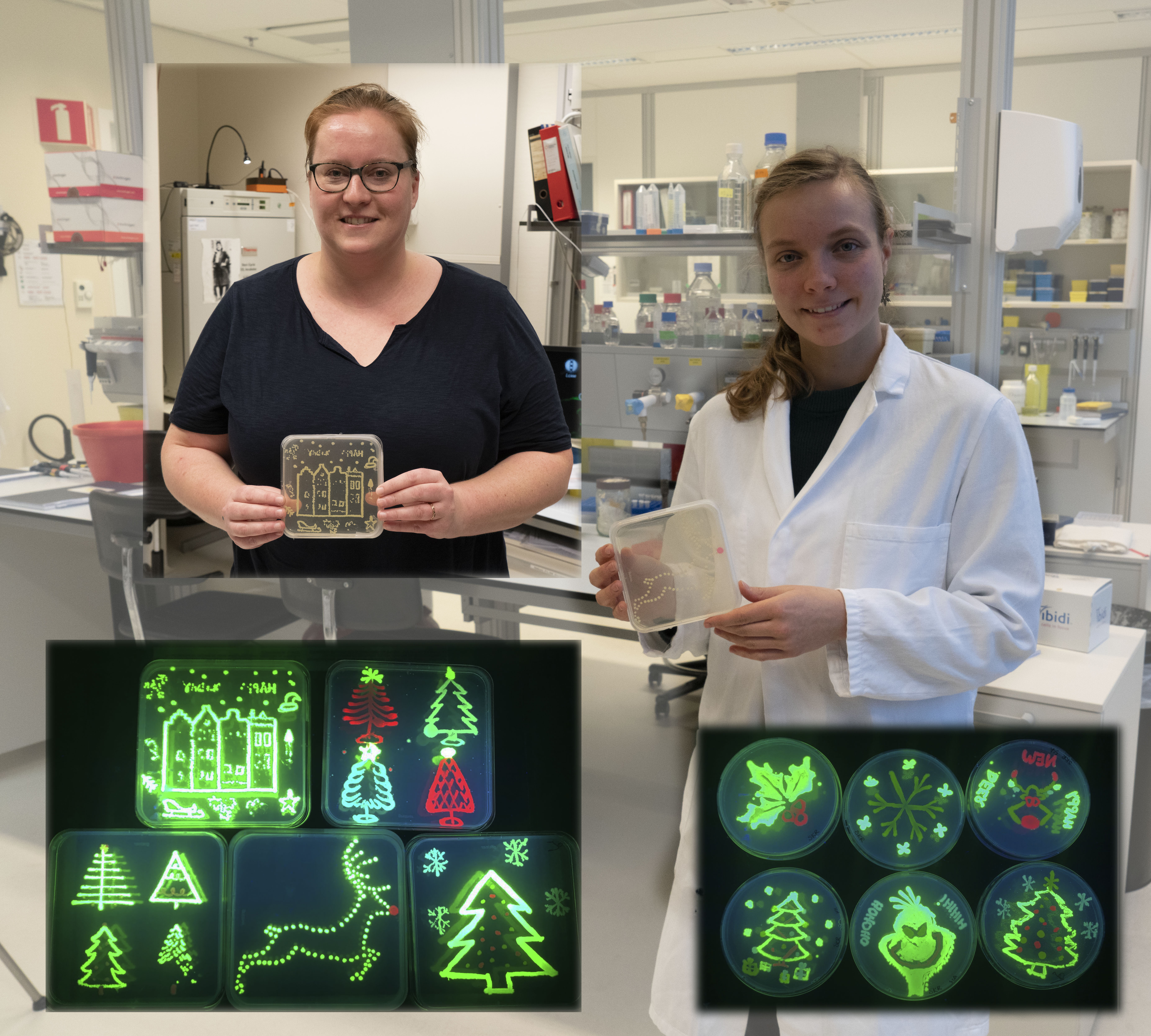
Assistant Professor Joachim Goedhart is a researcher who is developing these colored variants of the GFP protein at the University of Amsterdam in The Netherlands. I had the pleasure of interviewing him at his lab, where he showed us E. coli cultures which were transformed with GFP variant plasmids, resulting in spectacular colored bacterial colonies that are viewed under UV light. Joachim hopes that his plasmids help other researchers in visualizing their favorite protein, and deposits the sequences at Addgene, a global, nonprofit plasmid repository.
References:
- Green fluorescent protein – a bright idea for the study of bacterial protein localization (Gregory J Phillips) FEMS Microbiology Letters, Volume 204, Issue 1, 1 October 2001, Pages 9–18 (https://doi.org/10.1111/j.1574-6968.2001.tb10854.x)
- https://www.nobelprize.org/prizes/chemistry/2008/illustrated-information/
- Improved green flourescence (Heim R, Cubitt AB, Tsien RY) Nature. 373 (6516): 663–4 (https://doi.org/10.1038/373663b0)

This work is licensed under a Creative Commons Attribution-NonCommercial 4.0 International License.
About The Author
Sheba AJ
Sheba is the founder of WeTalkScience.com. Find her on Twitter at https://twitter.com/ShebaAJ
More about light (and the colours it hides…): https://www.space.com/25382-cosmos-recap-hidden-light-mysteries.html
Congratulations for this beautiful initiative, meaningful and important!
All the best and good luck!!, looking forward for more….
Thank you 🙂
Thanks. That was interesting